Understanding dizziness, migraine and persistent vestibular symptoms
One of the most distressing moments for people living with ongoing dizziness, imbalance or vertigo is hearing the words:
“Your scans are normal.”
On the surface, this sounds like good news. No tumour. No stroke. No structural damage. But for many people, it lands very differently. If your symptoms are persistent, debilitating and affecting daily life, a normal scan can feel invalidating — as though the problem must be imagined, exaggerated or “just anxiety.”
Nothing could be further from the truth.
In vestibular conditions such as vestibular migraine, persistent postural-perceptual dizziness (PPPD) and vestibular neuritis, normal scans are not only common — they are expected. These conditions are real, biological disorders of how the brain and inner ear function, not how they look on standard imaging.
Understanding this distinction is often a turning point in recovery.
What scans are actually designed to detect
To understand why scans are frequently normal in vestibular disorders, it helps to clarify what common investigations are — and are not — designed to show.
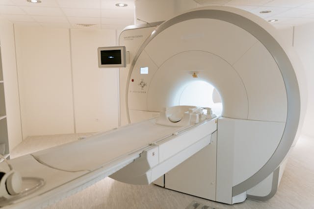
MRI and CT scans are excellent at detecting structural abnormalities. They can identify things like bleeding, tumours, large strokes, fractures, significant inflammation or obvious anatomical damage. They are lifesaving tools when used for the right indications.
What they do not measure well is function.
They do not show how accurately the inner ear is sending signals. They do not show how efficiently the brain is integrating balance information. They do not capture subtle changes in neural firing, sensory weighting, prediction error or autonomic regulation. And they do not reflect how the nervous system behaves under movement, fatigue, stress or visual load.
Vestibular symptoms almost always arise from problems in information processing, not from visible lesions.
The vestibular system is a prediction system, not a static structure
Balance is not a passive sense. Your brain is constantly predicting where your head and body should be in space, comparing that prediction with incoming sensory information from the inner ear, eyes and body, and making rapid corrections when something doesn’t match.
Dizziness arises when there is a mismatch, not necessarily damage.
If the signals don’t line up — even subtly — the brain generates symptoms to alert you that something isn’t right. Those symptoms may include vertigo, rocking, swaying, nausea, visual sensitivity, brain fog or fatigue.
Crucially, none of this requires anything to look abnormal on a scan.
Vestibular neuritis: when the damage is microscopic, not visible
Vestibular neuritis is a condition where the vestibular nerve — the nerve carrying balance information from the inner ear to the brain — becomes inflamed, often following a viral illness.
During the acute phase, people experience severe vertigo, nausea and imbalance. Over time, the inflammation settles, but the nerve does not always return to its previous level of function.
Here’s the key point: that nerve injury is often microscopic.
MRI scans are not sensitive enough to detect subtle changes in nerve firing, partial signal loss or altered timing. By the time scans are performed, the inflammation has often resolved, leaving behind a functional deficit rather than visible damage.
The brain must then recalibrate to this new input. If compensation is incomplete — or disrupted by inactivity, visual dependence or anxiety — ongoing dizziness can persist long after the scan appears “normal.”
The symptoms are real. The injury happened. It just isn’t something imaging can reliably show. A Vestibular Physiotherapist or Vestibular Audiologist will know how to test this more efficiently with a head impulse test or Vestibular Function test, not an MRI.
Vestibular migraine: a disorder of brain processing, not structure
Vestibular migraine is one of the most misunderstood causes of dizziness. Many people assume migraine must involve headache, or that something dramatic must show up on imaging. In reality, vestibular migraine is a neurological disorder of sensory processing.
In vestibular migraine, the brain becomes overly sensitive to normal sensory input. Signals that would normally be filtered out are amplified. The threshold for triggering symptoms drops. Visual motion, head movement, hormonal changes, sleep disruption, stress or dietary factors can all tip the system into overload.
During an attack, the brain’s balance networks behave differently — but they return to baseline between episodes. There is no tumour. No lesion. No permanent structural change to capture on a scan.
This is why imaging is typically normal, even in people with severe, recurrent symptoms. Occasionally there may be “white matter hyperintensities” in Migraine sufferers, however this is not overly common.
A normal scan does not rule out vestibular migraine. It supports it.
PPPD: when the brain stays in “high alert” mode
Persistent postural-perceptual dizziness (PPPD) often develops after a triggering event such as vestibular neuritis, BPPV, concussion, panic attacks or vestibular migraine.
In PPPD, the initial injury or episode resolves, but the brain remains stuck in a protective, hyper-vigilant state. Balance becomes consciously monitored instead of automatic. Vision becomes over-weighted. Movement is interpreted as threatening. Symptoms persist daily or near-daily.
This is not psychological in the way people often fear. It is neurobiological.
The brain’s threat and balance networks remain tightly coupled, even when the original trigger has passed. Standard imaging cannot show this persistent state of altered sensory integration or autonomic arousal.
Once again, the scan is normal because the hardware is intact — the issue lies in how the system is being run.
Why “normal” scans are often reassuring — even if they don’t feel that way
It’s understandable to feel frustrated or dismissed when tests come back clear. But in vestibular medicine, normal imaging often carries important meaning.
It tells us there is no progressive or dangerous structural disease. It rules out conditions that require urgent or invasive treatment. And it allows clinicians to confidently focus on functional recovery, rather than searching endlessly for something that isn’t there.
In many cases, a normal scan helps narrow the diagnosis rather than exclude it.
Why symptoms can feel severe even when scans are normal
People often struggle with the mismatch between how intense their symptoms feel and how “unremarkable” their test results look.
This happens because the vestibular system is deeply connected to survival circuits in the brain. Disruption to balance triggers nausea, fear, autonomic symptoms and a powerful sense of threat. Even small errors in processing can produce outsized symptoms.
Think of it like a smoke alarm. A small amount of smoke can produce a very loud response — not because the fire is large, but because the system is designed to err on the side of caution.
Vestibular symptoms are similar. The intensity of symptoms does not correlate with the visibility of damage on imaging.
The danger of misinterpreting normal scans
When people are told “everything is normal” without explanation, several unhelpful things can happen.
Some people are left searching endlessly for new tests, hoping something will finally “show up.” Others begin to doubt their own experience or blame themselves. Some are told their symptoms are anxiety-based and stop seeking appropriate vestibular care.
None of these pathways support recovery.
Understanding that vestibular conditions are diagnosed clinically, based on history, symptom patterns and examination — not solely on imaging — is crucial.
How vestibular rehabilitation fits into this picture
Because these conditions are functional rather than structural, recovery focuses on retraining the system, not fixing something visible on a scan.
Vestibular rehabilitation helps the brain recalibrate how it processes balance, motion and visual information. It reduces visual dependence, restores automatic balance control and gradually lowers the nervous system’s threat response to movement.
In vestibular neuritis, rehabilitation supports central compensation for reduced nerve input. In vestibular migraine, it improves motion tolerance and reduces sensory sensitivity between attacks. In PPPD, it helps break the cycle of hypervigilance and avoidance that keeps symptoms going.
This is why people often improve dramatically with the right rehabilitation program even after months or years of “normal” test results.
A reframing that matters
A normal scan does not mean:
- nothing is wrong
- symptoms are imagined
- recovery isn’t possible
It means:
- the structure is intact
- the issue lies in function, integration and regulation
- the nervous system can change
And that last point is the most important of all.
The brain is adaptable. With the right input, it can relearn safety, recalibrate balance and reduce symptoms — even when scans never change.
The takeaway
Vestibular conditions such as vestibular migraine, PPPD and vestibular neuritis are real, biological disorders that commonly occur without abnormalities on imaging. They affect how the brain and inner ear communicate, not how they look on a scan.
Normal investigations are not a dead end. They are often the starting point for the most effective treatment approach.
If you have persistent dizziness, imbalance or motion sensitivity despite normal scans, you are not alone — and you are not out of options. With accurate diagnosis, education and targeted vestibular rehabilitation, recovery is not only possible, it is expected.